Definition
By: Gregory R. Waryasz, MD
Copyright 2011
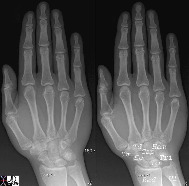
Normal Hand |
| 45737c01 hand wrist phalanx phalanges radius ulna wrist carpals carpal bones scaphoid lunate triquetrum pisiform hamate hook of hamate capitate tapezoid trapezium metacarpals metacarpal bone sesamoid normal anatomy plain film Courtesy Ashley Davidoff MD 45737 45738 45739 45741 |
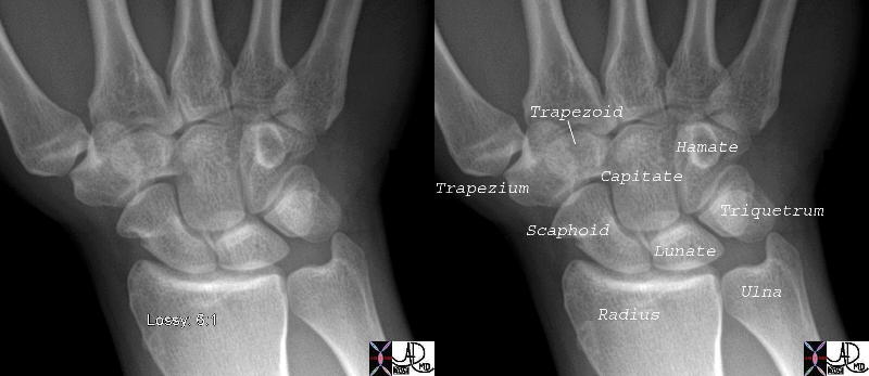
Normal Wrist |
| 45738c01 bone hand wrist radius ulna wrist carpals carpal bones scaphoid lunate triquetrum pisiform hamate hook of hamate capitate tapezoid trapezium metacarpals metacarpal bone normal anatomy plain film Courtesy Ashley Davidoff MD 45737 45738 45739 45741 |

CT Wrist |
| 46616b01 bone hand carpals wrist triquetral lunate scaphoid hamate capitate trapezoid trapezium radius ulna normal anatomy normal CTscan Davidoff MD |
|
Normal Wrist |
| 46613c01.800 bone hand carpals wrist hook of hamate shape anatomy normal CTscan Davidoff MD |
Applied Biology
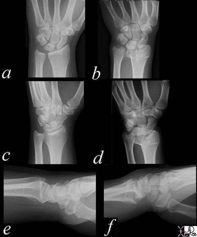
Normal and Colle’s Fracture |
| 70034c03 bone radius ulna wrist dorsally angulated fracture of distal radial metaphysis 2-3 cm proximal to wrist joint,sometimes associated frx of ulnar styloid frx line is almost always on volar side dx Colle’s fracture plain X-ray plain film of wrist normal wrist 70034c03 70034c04 Davidoff MD |
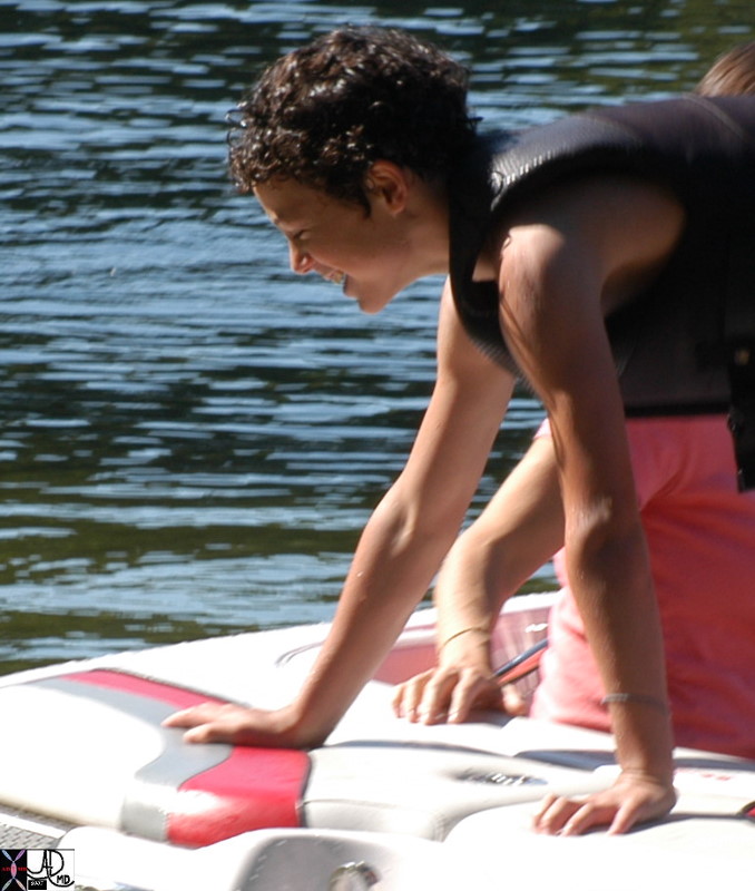
Mechanism for Colle’s Fracture |
| 85312p.800 falling outstetched arm Colle’s fracture mechanism of injury bone fracture Davidoff photography Davidoff MD |
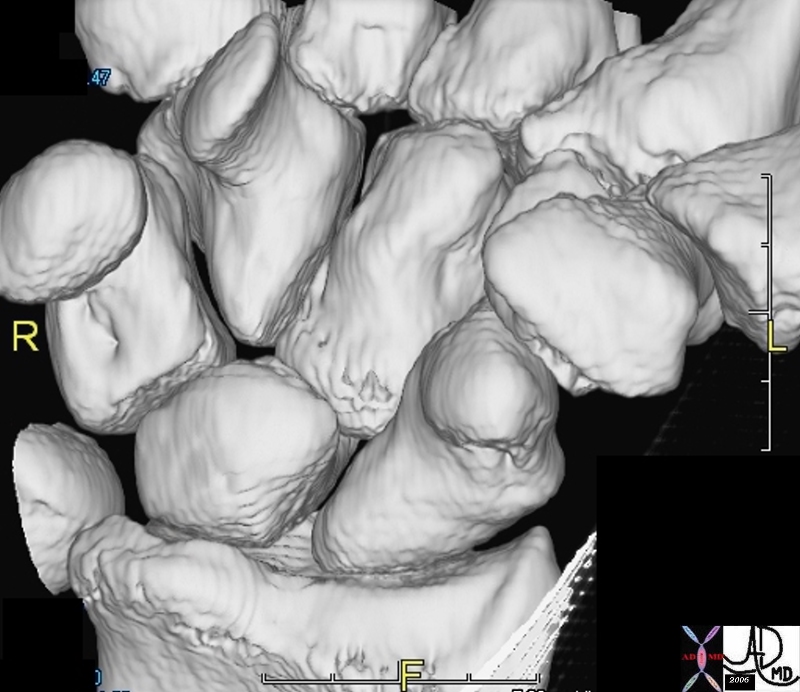
Bones of the Wrist |
| 46613 bone hand carpals wrist triquetral lunate scaphoid hamate capitate trapezoid trapezium radius ulna normal anatomy normal CTscan Davidoff MD |
The wrist joint of the musculoskeletal system is characterized by connecting the hand and the forearm.
It is part of the upper extremity and consists of bone, skeletal muscle, cartilage, synovial tissue, and tendon.
Its unique structural is that it is made up of a pivot joint and a condyloid type of synovial joint. The pivot joint is the distal radioulnar joint. The condyloid type of synovial joint is the radiocarpal joint.
The bones are the radius, ulna, and 8 carpal bones (scaphoid, lunate, triquetrum, pisiform, trapezium, trapezoid, capitate, and hamate). The articular surfaces are covered with cartilage.
The ligaments are the anterior and posterior ligament of the distal radioulnar joint, radial collateral ligament of the wrist, ulnar collateral ligament of the wrist, dorsal radiocarpal ligaments, and the palmar radiocarpal ligaments.
There is a fibrocartilaginous articular disc within the distal radioulnar joint. The triangular fibrocartilaginous complex is a central meniscus homologue.
The blood supply is from the anterior and posterior interosseous arteries to the distal radioulnar joint. The blood supply is from the branches of the dorsal and palmar carpal arches.
The innervation is from the anterior and posterior interosseous nerves to the distal radioulnar joint. The innervation of the radiocarpal joint is from the anterior and posterior interosseous nerves and the ulnar nerve.
The wrist joint’s components as well as all other bones, muscles, and ligaments of the body are derived of mesodermal origin in the embryo.
The function of the wrist is to allow for pronation/supination, flexion/extension, and abduction/adduction.
Supination occurs by the supinator and the biceps brachii. There is some supination from the extensor pollicis longus and the extensor carpi radialis longus. The radius uncrosses form the ulna by the distal radius rotating laterally and posteriorly to make the bones into parallel arrangement.
Pronation occurs from the pronator quadrates and the pronator teres. Other muscles that assist with pronation include the flexor carpi radialis, palmaris longus, and brachioradialis. During pronation, the distal radius rotates anteriorly and medially crossing over the ulna anteriorly.
Flexion of the wrist is by the flexor carpi radialis and flexor carpi ulnaris. There is some assistance from the flexors of the thumb and fingers, palmaris longus, and the abductor pollicis longus.
Extension of the wrist is by the extensor carpi radialis longus, extensor carpi radialis brevis, and extensor carpi ulnaris. There is some assistance from the finger extensor and the thumb.
Abduction of the wrist is by the abductor pollicis longus, flexor carpi radialis, extensor carpi radialis brevis, and extensor carpi radialis longus.
Adduction of the wrist is by the simultaneous contraction of the extensor carpi ulnaris and the flexor carpi ulnaris.
Common diseases of the wrist joint include fracture, dislocation, tendinitis, arthritis, septic arthritis, transient synovitis, ligament injury, triangular fibrocartilagenous complex (TFCC) tear, carpal tunnel syndrome, Kienbock’s disease, and ganglion cysts.
Fractures can occur in the carpal, radial, or ulnar components of the wrist joint. There can be an associated dislocation with the fracture.
Dislocation without fracture can also occur.
Osteoarthritis is a condition of joint space narrowing leading to pain. Rheumatoid arthritis is a condition of an autoimmune reaction against the synovial tissue.
Tendinitis is a condition of overuse resulting in inflammation of the tendon.
Tenosynovitis is inflammation of the synovial tendon sheath. It commonly occurs at with the abductor pollicis longus, extensor pollicis brevis, and flexor carpi radialis. These lead to pain in the wrist. De quervain tenosynovitis is specifically either the abductor pollicis longus and/or extensor pollicis brevis tendons.
Ligament injuries can occur including full thickness or partial tears.
Tears can occur in the triangular fibrocartilagenous complex (TFCC).
Transient synovitis or toxic synovitis is joint pain after a recent viral illness or trauma. It is a self-limiting condition of patients between the ages 3 to 8.
Lyme Arthritis is a condition common in the northeast USA where there is a rheumatologic reaction to Borrelia burgdoferi. It occurs after the patient is bitten by a tick.
Osteomyelitis is an infection of the bone typically caused by a bacteria.
Osteoporosis is a condition of low bone mineral density that can predispose an individual to fractures.
Septic arthritis is an infection of the synovial tissue of the joint with pus in the joint cavity. The infection is capable of rapidly destroying the joint.
Carpal tunnel syndrome is a compressive neuropathy of the median nerve leading to weakness in the hand and numbness/tingling in the distribution of the median nerve.
Kienbock’s disease is a condition of avascular necrosis of the lunate. There is collapse of the lunate bone and in later stages fracture. It can become very painful and limit wrist range of motion.
A ganglion cyst is a mucinous filled cyst near the joint capsule or tendon sheath that occurs more commonly on the dorsal surface of the upper extremity.
Commonly used diagnostic procedures include clinical history, physical exam, x-ray, CT, EMG, and MRI.
It is usually treated with rest, NSAIDs, physical therapy, and possibly surgery. Dislocation/subluxation and fractures may not respond to nonoperative treatments and may require surgery. Arthritis can be treated with NSAIDs and physical therapy, but may require surgery. Tendinitis and tenosynovitis are treated with rest, NSAIDs, physical therapy, and corticosteroid injections. Septic arthritis requires surgical washout and antibiotics. Transient synovitis is treated with NSAIDs. Ligament or TFCC tears may require surgical repair or reconstruction. Kienbock’s disease can be initially treated non-operatively, but with advanced destruction of the lunate, may require surgery. Carpal tunnel syndrome is treated non-operatively with NSAIDs, physical therapy, and splints, but may require a carpal tunnel release surgery.
References
Davis MF, Davis PF, Ross DS. Expert Guide to Sports Medicine. ACP Series, 2005.
Elstrom J, Virkus W, Pankovich (eds), Handbook of Fractures (3rd edition), McGraw Hill, New York, NY, 2006.
Koval K, Zuckerman J (eds), Handbook of Fractures (3rd edition), Lippincott Williams & Wilkins, Philadelphia, PA, 2006.
Lieberman J (ed), AAOS Comprehensive Orthopaedic Review, American Academy of Orthopaedic Surgeons, 2008.
Moore K, Dalley A (eds), Clinically Oriented Anatomy (5th edition), Lippincott Williams & Wilkins, Philadelphia, PA, 2006.
Wheeless’ Textbook of Orthopaedics: Wrist Joint Menu (http://www.wheelessonline.com/ortho/wrist_menu)

