
Bones

| Sternum Trees |
| 46650b07b01.800 bone sternum costal cartilages calcified Davidoff art Davidoff Trees |

| Sternum Trees |
| 46650b22.800 bone sternum costal cartilages calcified river mountains sun water Davidoff |

Sternum Trees |
| 46650c01 bone sternum costal cartilages calcified Davidoff trees Davidoff art |
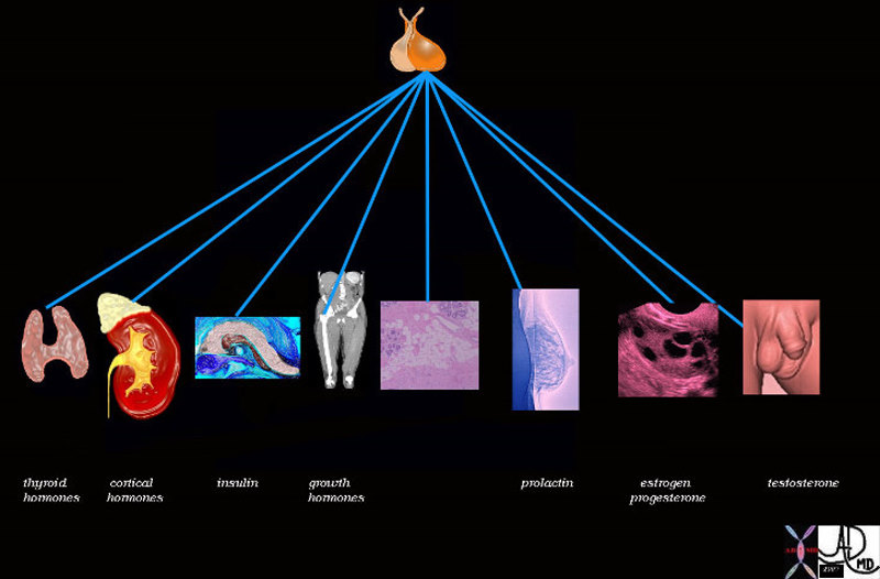
Pituitary Control |
| 72353 pituitary gland posterior pituitary gland thyroid adrenal cortex pancreas insulin bone muscle growth adipose tissue fat glucose parathyroid gland calcium breast lactation prolactin ovary estrogen FSH progesterone testis testes testosterone function physiology specialised function control endocrine system Davidoff art Davidoff drawing Davidoff MD |

Bones of the Wrist |
| 46613 bone hand carpals wrist triquetral lunate scaphoid hamate capitate trapezoid trapezium radius ulna normal anatomy normal CTscan Davidoff MD |
The Osteon – Unit of Compact Bone |
|
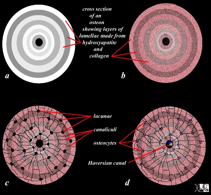
The diagram shows the microscopic appearance of compact bone with columnar morphology of the individual unit of compact bone called the osteon (overlaid in red) The osteon (aka Haversian syatem) is about 2 mms. in diameter It consists of concentric layers or lamellae that surround the Haversian canal that contains blood vessels (red and blue ) and nerves CODE bone osteon Haversian system Haversian canal compact bone cortex bone cortical bone lamella lamellae anatomy normal principles diagram Awaiting Permission fpr Use Image Rendered by Ashley Davidoff 106272b06b01.8 |
The OSteon in Axial Section showing the Lamellae |
|
The diagram illustrates the appearance of an osteon (Haversian system) in cross section. The osteon is the unit or building brick of compact bone and consists of columns of bone with layered subunits called lamellae. The lamellae consist of calcium, phosphate, and hydroxyl ions which form a compound called hydroxyapatite (Ca5(PO4)3(OH) – seen in (a) as shades of white and gray overlay. The hydroxyapatite gives bone its hardness. The lamella also contains type I collagen fibers. (color overlay of reds and pinks on the white background). The collagen provides elasticity. Lacunae (c) are spaces in the lamellae that house the osteocytes (d). The canaliculi(c) are a network of fine tubular channels that allow the osteocytes to communicate with each other. The Haversian canal (c,d) spaces in the middle of the osteon that contains artery, vein and nerve (c). Courtesy Ashley Davidoff Copyright 2011 106275c03L08b09cLd02.81s |

Columnar Nature of the Osteon |
|
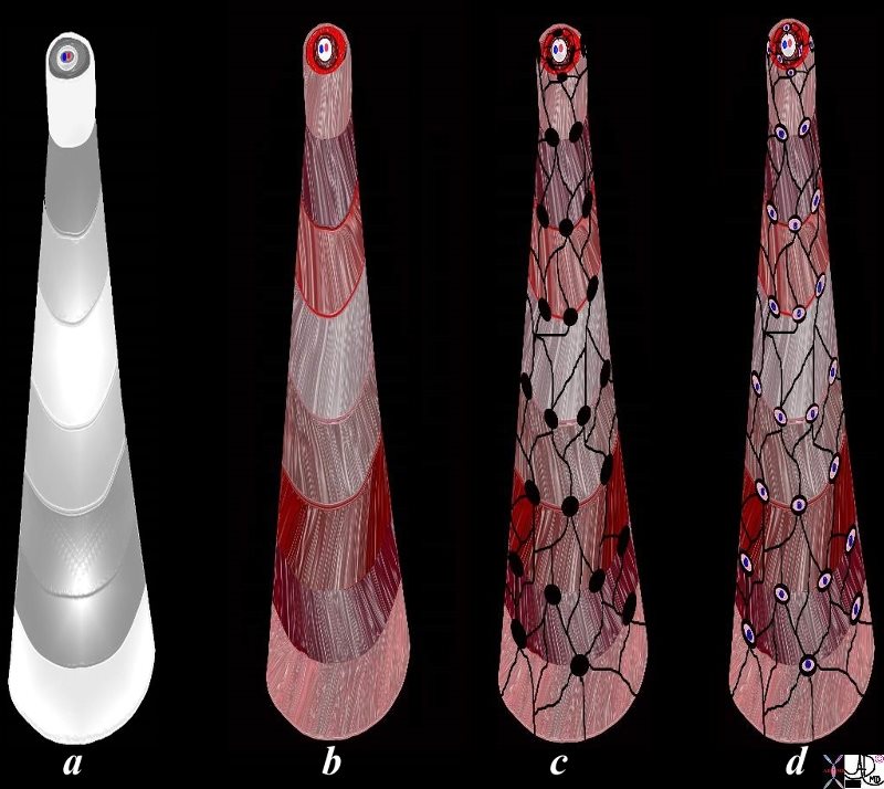
The diagram illustrates the appearance of an osteon (Haversian system) in 3D. The osteon is the unit or building brick of compact bone and consists of a column of bone with layered subunits called lamellae. The lamellae consist of calcium, phosphate, and hydroxyl ions which form a compound called hydroxyapatite (Ca5(PO4)3(OH) – seen in (a) as shades of white and gray overlay. The hydroxyapatite gives bone its hardness. The lamella also contains type I collagen fibers. (color overlay of reds and pinks on the white background) that are arranged in different directions on adjacent lamellae so that a zig zag pattern of the collagen is created allowing for strentheniing of the tissue. The collagen provides elasticity. Lacunae (c) are spaces in the lamellae that house the osteocytes (d). The canaliculi(c) are a network of fine tubular channels that allow the osteocytes to communicate with each other. The Haversian canal (c,d) spaces in the middle of the osteon that contains artery, vein and nerve (c). Courtesy Ashley Davidoff MD 106275c03L11.43kce02L.8s |
The Osteon and its Haversian Canal, Lamellae Lacunae, Canaliculi and Osteocytes |
|
The diagram illustrates the appearance of an osteon (Haversian system) in 3D. The osteon is the unit or building brick of compact bone and consists of a column of bone with layered subunits called lamellae. In this artistic rendering the inner most lamella has been pulled upward, dragging and stretching the sequential lamellae so as to display the morphology of all the lamellae in the osteon. The lamellae consist of calcium, phosphate, and hydroxyl ions which form a compound called hydroxyapatite (Ca5(PO4)3(OH) – seen in (a) as shades of white and gray overlay. The hydroxyapatite gives bone its hardness. The lamella also contains type I collagen fibers. (color overlay of reds and pinks on the white background) that are arranged in different directions on adjacent lamellae so that a zig zag pattern of the collagen is created allowing for strengthening of the tissue. The collagen provides elasticity. Lacunae (c, black holes) are spaces in the lamellae that house the osteocytes (d). The canaliculi(c black web) are a network of fine tubular channels that allow the osteocytes to communicate with each other. The Haversian canal is the space seen on the top and in the middle of the osteons that contains artery, vein and nerve (a-d). Courtesy Ashley Davidoff Copyright 2011 106275c03L13.8s |
Slime – What is the Connection to the Bone? |
|
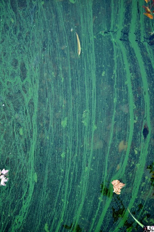
Slime on the surface of Crystal lake in Newton Massachusetts was reminiscent of the trabecular pattern of bone with both linear elements and spongy elements. The picture was taken in early Fall with fallen leaves having a free ride on the slime on the surface of the lake. The push and pull forces of the water on the slime created this pattern. Courtesy Ashley DAvidoff Md copyright 2011 107541pb.8 |
More Beauty in Slime |
|
Slime on the surface of Crystal lake in Newton Massachusetts was reminiscent of the trabecular pattern of bone with both linear elements and spongy elements. The picture was taken in early Fall with fallen leaves having a free ride on the slime on the surface of the lake. The push and pull forces of the water on the slime created this pattern. Courtesy Ashley DAvidoff Md copyright 2011 107542pb.8s |
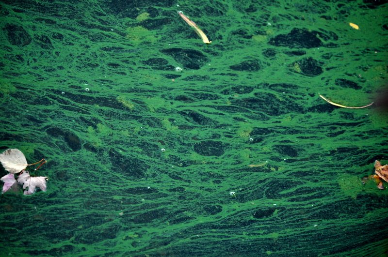
The Trabecular PAttern and Slime on the Lake |
|
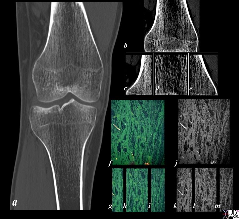
Slime on the surface of Crystal lake in Newton Massachusetts was reminiscent of the trabecular pattern of bone with both linear elements and spongy elements. The picture was taken in early Fall with fallen leaves having a free ride on the slime on the surface of the lake. The push and pull forces of the water on the slime created this pattern. The trabecular pattern of the bone in the tibia (a) is enhanced in b c d and e with the linear trabeculations noted on the medial amnd lateral aspect of the femur (c and e) while the spongy appearance is seen in the middle ((d). The appearance of the slime on the lake was reminiscent of this pattern (f) and divided into the lateral (g) and medical (i) regions with linear patterns and the middle with spongy appearance (h) The image in j k l and m represent the black and white of the slime image Courtesy Ashley Davidoff MD copyright 2011 107570pcc01L.1kb.8s |
Collagen Type 1 Fibers and Basketball Net |
|
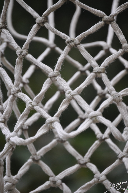
The basketball net has a zig zag shape that allows the net to be pliable but strong. The pattern of the collagen type 1 fibres in the lamellae of bone is also in a zig zag pattern affording the compact bone pliability some elasticity and strength as well. Courtesy Ashley Davidoff Copyright 2011 107546p.8s |
Copyright 2011
Introduction
Fractures
Fractures of the Body |
|
The artistic rendering of fractures of the skeletal system starts with the top row of images representing the skull and c-spine fractures, followed by thoracic and lumbar spinein the next row, and progressively moving down through the upper limbs, pelvis and lover limbs with the last row representing fractures of the feet Courtesy Ashley Davidoff MD copyright 2011 all rights reserved 88361c.8s |
Fractures – Pain |
|
The artistic rendering of fractures of the skeletal system are typified by a dramatic comminuted fracture of the skull that dominates the centre of the image. The pain of the patient is palpable. The top row then starts with images representing the skull and c-spine fractures, followed by thoracic and lumbar spinein the next row, and progressively moving down through the upper limbs, pelvis and lover limbs with the last row representing fractures of the feet Courtesy Ashley Davidoff MD copyright 2011 all rights reserved 88361c.81s |
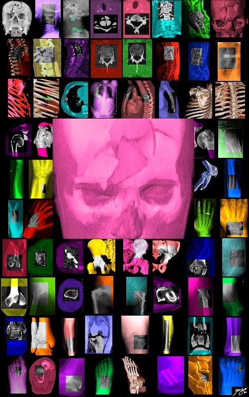
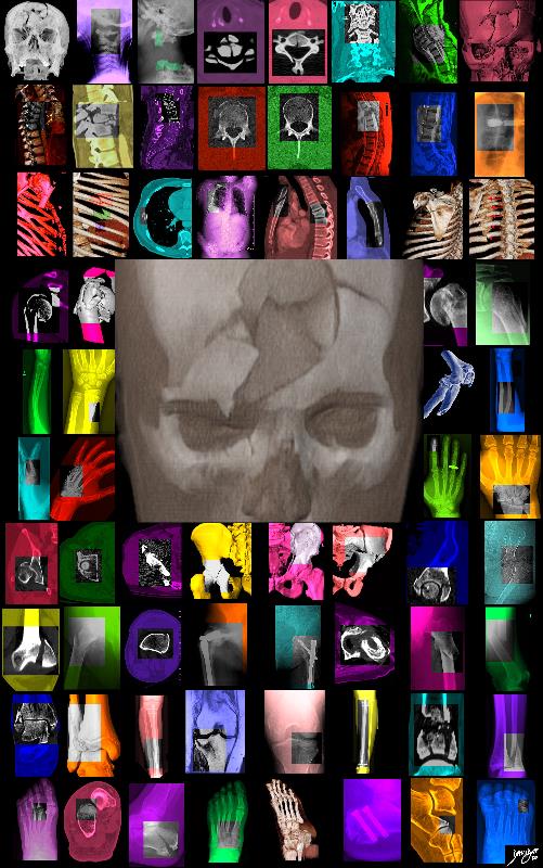
Skull Fracture |
|
The artistic rendering of fractures of the skeletal system are typified by a dramatic comminuted fracture of the skull that dominates the centre of the image. The pain of the patient is palpable. The top row then starts with images representing the skull and c-spine fractures, followed by thoracic and lumbar spinein the next row, and progressively moving down through the upper limbs, pelvis and lover limbs with the last row representing fractures of the feet Courtesy Ashley Davidoff MD copyright 2011 all rights reserved 88361c.82s |
|
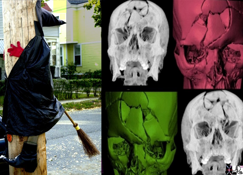
High Velocity Halloween Trauma |
|
A depressed comminuted fracture of the frontal bone often caused by a high speed decelerating injury with the patuients foreghead imapacting the steering wheel or front wind screen. In such an injury associated injuries such as whiplash injuries to the cervical spine or decelerating injury to the aorta can be life threatening. Courtesy Ashley Davidoff MD Copyright 2011 65840c04.8s Courtesy Ashley Davidoff MD Copyright 2011 65840.8s |
Structural Principles
Compact Bone
|
The Osteon with its Haversian Canal, Lamellae, Lacunae, Canaliculi and Osteocytes |
|
The diagram illustrates the appearance of an osteon (Haversian system) in 3D. The osteon is the unit or building brick of compact bone and consists of a column of bone with layered subunits called lamellae. In this artistic rendering the inner most lamella has been pulled upward, dragging and stretching the sequential lamellae so as to display the morphology of all the lamellae in the osteon. The lamellae consist of calcium, phosphate, and hydroxyl ions which form a compound called hydroxyapatite (Ca5(PO4)3(OH) – seen in (a) as shades of white and gray overlay. The hydroxyapatite gives bone its hardness. The lamella also contains type I collagen fibers. (color overlay of reds and pinks on the white background) that are arranged in different directions on adjacent lamellae so that a zig zag pattern of the collagen is created allowing for strengthening of the tissue. The collagen provides elasticity. Lacunae (c, black holes) are spaces in the lamellae that house the osteocytes (d). The canaliculi(c black web) are a network of fine tubular channels that allow the osteocytes to communicate with each other. The Haversian canal is the space seen on the top and in the middle of the osteons that contains artery, vein and nerve (a-d). The diagram illustrates the appearance of an osteon (Haversian system) in 3D. The osteon is the unit or building brick of compact bone and consists of a column of bone with layered subunits called lamellae. In this artistic rendering the inner most lamella have been pulled upward so as to display the morphology of all the lamellae. The lamellae consist of calcium, phosphate, and hydroxyl ions which form a compound called hydroxyapatite (Ca5(PO4)3(OH) -. The hydroxyapatite gives bone its hardness. The lamella also contains type I collagen fibers. (color overlay of reds and pinks) that are arranged in different directions on adjacent lamellae so that a zig zag pattern of the collagen is created allowing for strengthening of the tissue (a). The collagen provides elasticity. Lacunae (b) are spaces in the lamellae that house the osteocytes (c). The canaliculi are a network of fine tubular channels that allow the osteocytes to communicate with each other. The Haversian canal spaces in the middle of the osteon that contains artery, vein and nerve. Courtesy Ashley Davidoff Copyright 2011 106275c03L13.8s |
Trabecular Bone
|
Trabecular or Spongy Pattern of Slime on the Lake |
|
Slime on the surface of Crystal lake in Newton Massachusetts was reminiscent of the trabecular pattern of bone with both linear elements and spongy elements. The picture was taken in early Fall with fallen leaves having a free ride on the slime on the surface of the lake. The push and pull forces of the water on the slime created this pattern. Courtesy Ashley DAvidoff Md copyright 2011 107541pb.8 |
|
More beauty in Slime |
|
Slime on the surface of Crystal lake in Newton Massachusetts was reminiscent of the trabecular pattern of bone with both linear elements and spongy elements. The picture was taken in early Fall with fallen leaves having a free ride on the slime on the surface of the lake. The push and pull forces of the water on the slime created this pattern. Courtesy Ashley Davidoff Md copyright 2011 107542pb.8s |
|
The Trabecula and The Pattern of Slime on the Lake |
|
Slime on the surface of Crystal lake in Newton Massachusetts was reminiscent of the trabecular pattern of bone with both linear elements and spongy elements. The picture was taken in early Fall with fallen leaves having a free ride on the slime on the surface of the lake. The push and pull forces of the water on the slime created this pattern. The trabecular pattern of the bone in the tibia (a) is enhanced in b c d and e with the linear trabeculations noted on the medial amnd lateral aspect of the femur (c and e) while the spongy appearance is seen in the middle ((d). The appearance of the slime on the lake was reminiscent of this pattern (f) and divided into the lateral (g) and medical (i) regions with linear patterns and the middle with spongy appearance (h) The image in j k l and m represent the black and white of the slime image Courtesy Ashley Davidoff MD copyright 2011 107570pcc01L.1kb.8s |
|
Collagen Type 1 Fibers and the Basketball Net |
|
The basketball net has a zig zag shape that allows the net to be pliable but strong. The pattern of the collagen type 1 fibres in the lamellae of bone is also in a zig zag pattern affording the compact bone pliability some elasticity and strength as well. Courtesy Ashley Davidoff Copyright 2011 107546p.8s |
|
The Strong and the Weak – Bones and Logs |
|
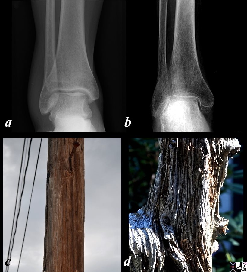
The images reflect an A-P examination of the ankle of a normal 21 year old male and the ankle of an 81 year old osteoporotic female (b) juxtaposed on two logs with similar morphological differences. In the osteoporotic female (b) the trabeculae of the bone are prominent and the overall density of the bone is decreased. The log in image c is in use as a telephone pole, sturdy enough to carry electrical and phone lines and hard enough to retain large metallic pegs that are sufficient in strength to maintain the weight of a full grown man. The attachments of pegs and wires are equivalent to the demands placed on a normal bone that has tendons, ligaments, and muscles attached. The second log (d) has lain on the ground detached from its mother tree, ill prepared for many years of disuse. The matrix has been lost and the “trabeculae” of the skeleton are prominent. This branch would be too weak to support the wires and large metallic pegs compared to the log in c primed when removed from the earth to function at its full strength. These 4 images project the difference between normal strength bone/log (a,c) and osteoporotic bone and weakened log (b,d). Osteoporosis is an entity where the bone becomes weakened by loss of component elements. The trabeculae become prominent and the overall density of the bone is decreased. The osteoporotic bone is at risk for pathological fracture. Courtesy Ashley Davidoff Copyright 2011 107482pc.8s.c01L |
Time and the Spine Kyphosis Couple |
|
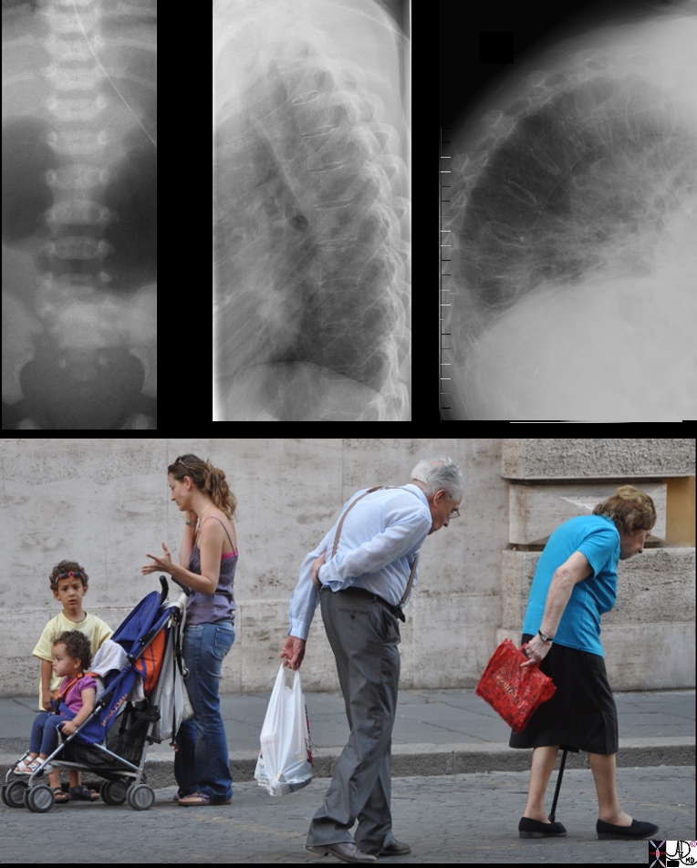
The top row of images from left to right reflect, A-P examination of the immature lumbar spine of very young patient, juxtaposed with the lateral examination of a normal thoracic of a normal young adult and lastly the lateral examination of a severely kyphotic elderly patient. The photograph was taken in Italy showing ages ranging from the youngest child in a stroller perhaps 2 years in age, her brother of about 5 or 6, their mother in her late twenties or early thirties and an elderly couple both suffering from the wraths of aging bones – osteoporosis and severe kyphosis. (the kyphosis couple) Courtesy Ashley Davidoff Copyright 2011 75578c01.8s |
| Sudden Force – The Way Many Fractures Happen
Compressive and Tensile Forces |
|
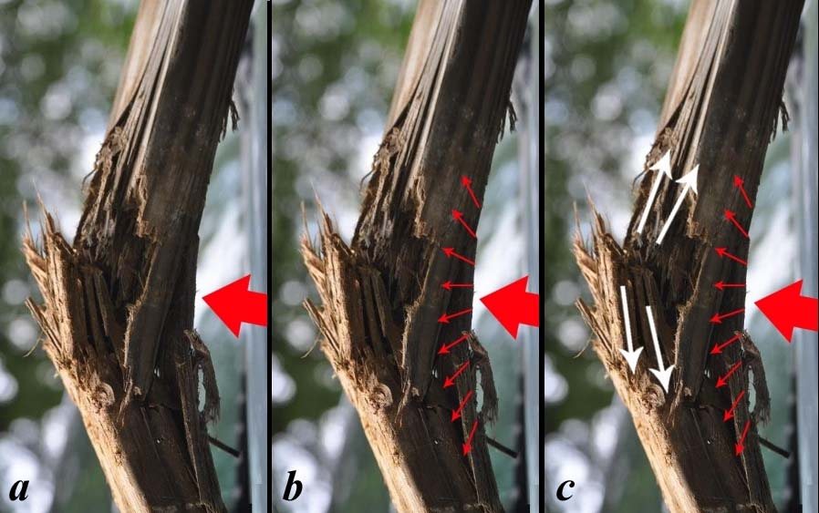
The series of images show the initial force on the bone (large red arrow in a) followed by the compressive force (small red arrows b) on the ipsilateral side of the injurious force. As the force gets transmitted through the bone tensile forces (white arrows in c) cause a distraction of the fibres of the bone.. When a pressure is applied to a long bone at an approximate right angle vector a pliable structure like bone will bend having a concave shape on the receiving end of the force and a convex shape on the opposite surface The forces on the concave side are compressive and on the opposite convexity they are tensile.(Latin tendere – to stretch). Courtesy Ashley Davidoff Copyright 2011 106647p.84cL.91L |
106647p.81 |
|
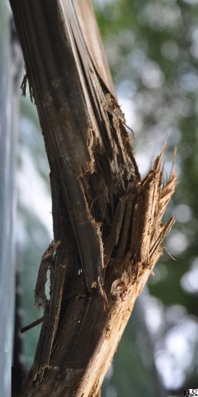
This photograph shows a splintered transverse fracture of a small branch of a tree showing a varus deformity. The fracture is not complete with the medial edge remaining intact. Courtesy Ashley Davidoff Copyright 2011 106647p.81 |
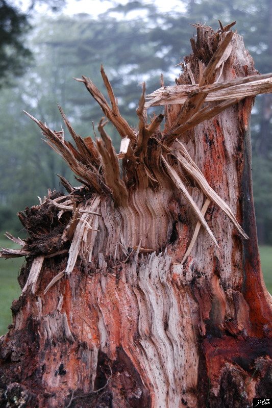
Splintered Fragments of a Fractured Trunk of a Tree |
| 87403p.8s tree Connecticut Berkshires fracture stump sharp pointed disease trauma Copyright 2009 all rights reserved Davidoff photography |
Macroscopic Insight into Microscopic Changes |
|
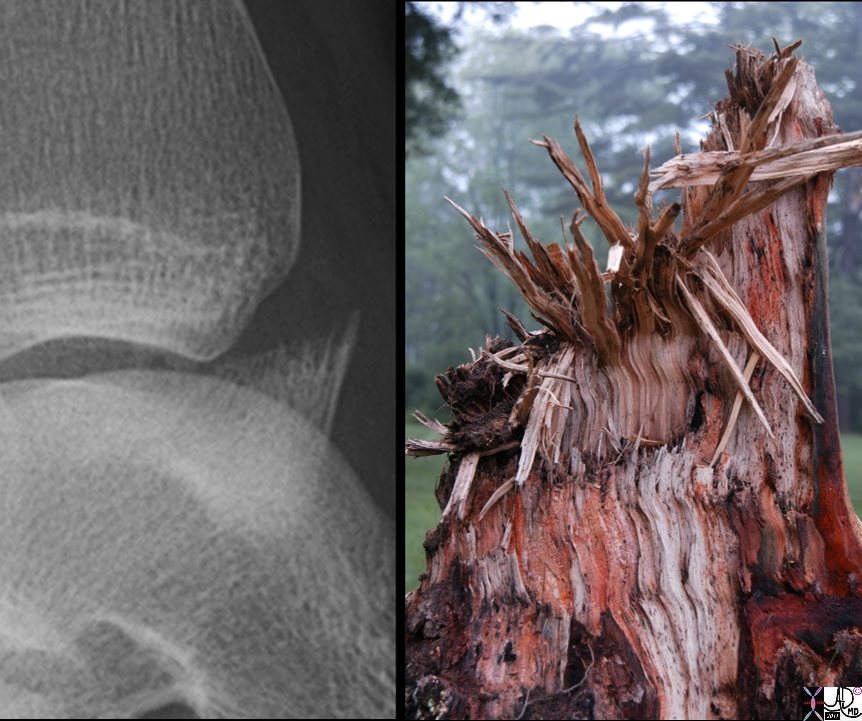
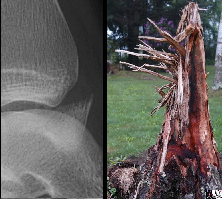
The X-ray of the ankle in a 15 year old male in the anteroposterior projection (A-P) shows an avulsion fracture of the anterior portion of the distal tibia. Note the spikes of broken bone at the edge of the distal fragment, reminiscent of the spikes of splintered tree from the weather induced fracture of this tree taken in the Berkshires Connecticut. The close relationship of the two bones in the ankle region and the presence of an interosseus membrane that binds the two bones into a single unit, makes combination fractures common. . Courtesy Ashley Davidoff Copyright 2011 99940c02 |
Macroscopic Insight into Microscopic Changes |
|
These images afford us the opportunity to get a sense of the shape and pathogenesis of the fracture at the fracture line because the roslution of the fracture is so clear in the X-ray image. The X-ray is of the ankle in a 15 year old male in the anteroposterior projection (A-P) and shows an avulsion fracture of the anterior portion of the distal tibia. Note the spikes of broken bone at the edge of the distal fragment, reminiscent of the spikes of splintered tree from the weather induced fracture of this tree. The photograph was taken in the Berkshires Connecticut. Courtesy Ashley Davidoff Copyright 2011 99940c02.8s |
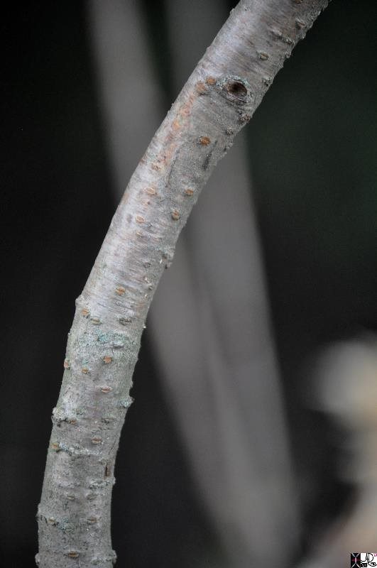
Bowing Injury a Type of Greenstick |
|
This photograph shows bowing of a small branch of a tree without any distinct fracture. This type of traumatic deformity is seen only in the pediatric population reflecting the softer and more pliable nature of bone. The intact innerconcave edge is the site of impact and the outer convex edge is on the opposite side of the impacting force. This image exemplifies a greenstick fracture. Courtesy Ashley Davidoff Copyright 2011 107345pb01.8 |
Fracture |
|
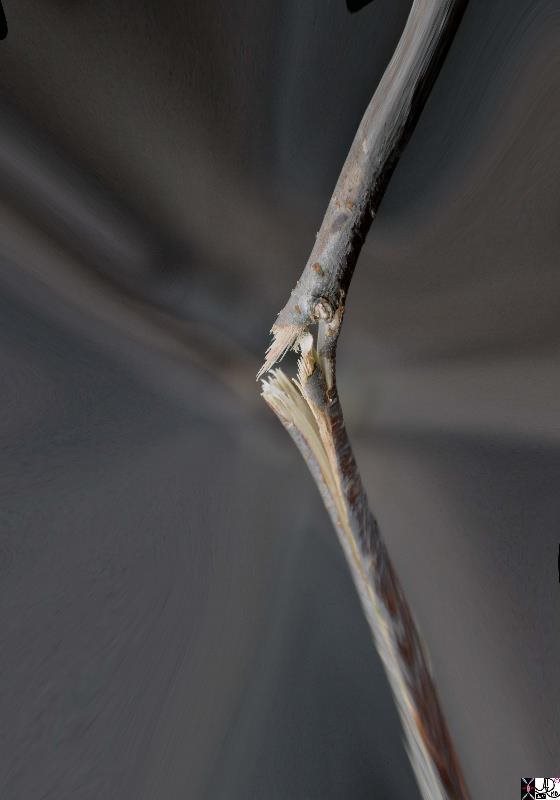
This photograph shows a splintered transverse fracture of a small branch of a tree showing a varus deformity if considered the right femur for example, but a valgus deformity if considered the left femur. The fracture is not complete with the medial edge (of a right sided fracture) remaining intact, held in place by the bark – the equivalent of the periosteum. The periosteum will remain intact on the side of the force while tearing occurs over the fracture on the opposite side. Courtesy Ashley Davidoff Copyright 2011 107342pb03.8 |
Greenstick Fracture |
|
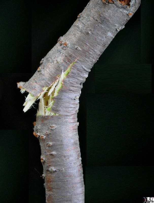
This photograph shows an incomplete splintered transverse fracture of a small branch of a tree The intact inner edge is usually the site of impact and the outer fractured edge edge is on the opposite. This image exemplifies a greenstick fracture characteristically seen in the pediatric population where bones are more pliable and less brittle. Courtesy Ashley Davidoff Copyright 2011 107344pb01.8 |
|
Complete Fracture Periosteum Intact |
|
This photograph shows a splintered transverse fracture of a small branch of a tree showing a varus deformity if considered the right femur for example, but a valgus deformity if considered the left femur. The fracture is not complete with the medial edge (of a right sided fracture) remaining intact, held in place by the bark – the equivalent of the periosteum. The periosteum will remain intact on the side of the force while tearing occurs over the fracture on the opposite side. Courtesy Ashley Davidoff Copyright 2011 107342pb03.8 |
Causes of Stress Fractures |
|
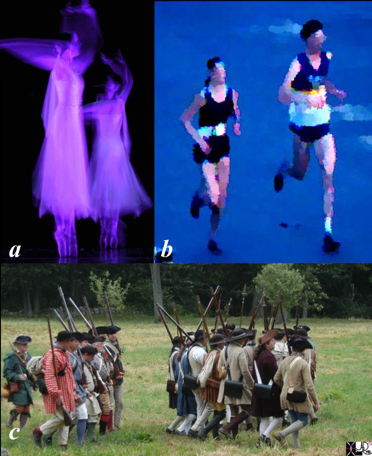
The 3 images project the activities that predispose to stress fractures. Image a shows two ballet dancers “en pointe” meaning that they are dancing on the tips of their toes with the full weight of the body transmitted through the toes, tarsals and metatarsals – a position that was not designed for all mortals. This takes immense skill and practice and predisposes to stress fractures. Image b shows two runners finishing the Boston marathon – after a 26 mile run. This takes immense talent and practice of repeated long distance and repetitive pounding on the back and legs. This activity also predisposes to stress fractures. Image c shows an army march in the fields of a reenacted battle of the revolution. Miles of marching predisposes to stress fractures of the lower legs and feet as well hence the name march fractures. Courtesy Ashley Davidoff Copyright 2011 84023ph01.8sL |
Amputation of the Tuft of the Finger and Fracture |
|
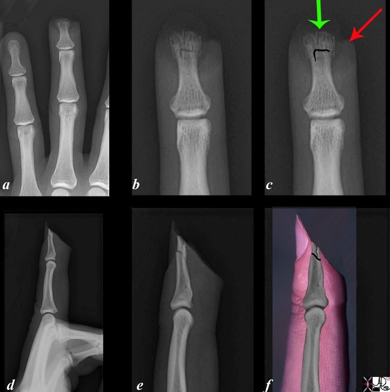
The series of X-ray are from a 29 year old man who amputated the tip of his right middle finger with a sharp power tool. Image a is an A-P view, magnified in image b and overlaid with the fracture line (black) in c. The green arrow points to the bone edge of the distal tuft, which is reduced in size and has lost its outer cortex, exposing a fragmented and trabeculated border. The red arrow points to the difficult to see skin edge. Image d is a lateral projection of the middle finger, magnified in image e, and overlaid with the photograph of the soft tissues of the finger and nail bed. This projection reveals the well defined and relatively straight skin and bone injury as a result of the sharpness of the instrument. The black line reveals the inverted “T” shaped fracture. Courtesy Ashley Davidoff Copyright 2011 102105bc07L.8s |
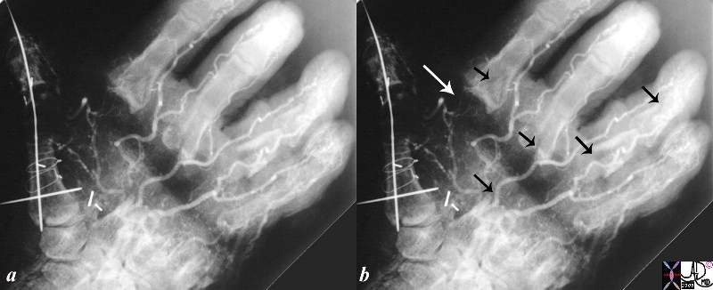
Fracture Horror! |
|
This is one of the most painful X-rays of my career and it is older than 30 years. The exact history eludes me but there has been obvious traumatic injury to the handwith multiple fractures (black arrows) a transverse skin incision that contains a broad band of air (white arrow), probable debridement of the severely injured metacarpal row, and extensive pinning and wiring of the thumb following surgical trauma. The combination of the large somatic sensory nociceptor supply to the hand, together with the extensive skin, bone,and tissue damage, is difficult to perceivethe extent of the pain thatthis poor patient went through. The remarkable feature is themaintenace of blood supply to the hand. Courtesy Ashley Davidoff MD copyright 2008 17496c02.8s |