Assistant
Definition
By Gregory R. Waryasz, MD
The radius of the musculoskeletal system is characterized by being the lateral and shorter bone in the forearm.
It is part of the upper limb. The radius is part of the elbow, wrist, and distal radial-ulna joints. It consists of bone and cartilage tissue once mature.
Its unique structural features include its gradual bend in the shaft. The radius extends from its articulation with the humerus to the carpus. The radius and ulna are connected by an interosseous membrane.
The proximal radius has a head with a central fovea and a neck. The proximal medial radial tuberosity is the site for insertion of the biceps tendon.
The radial shaft increases in diameter as it becomes distal. The radial shaft contains a long medullary cavity.
The distal radius has a carpal articular surface, an ulnar notch, a dorsal tubercle, and a lateral radial styloid process. The dorsal tubercle lies between grooves that allow for passage of the forearm muscle tendons. The radial styloid process is larger than the ulnar styloid and extends more distally.
The muscles that attach to the radius are the biceps brachii, supinator, flexor digitorum superficialis, flexor pollicis longus, pronator teres, pronator quadrates, and the supinator.
The radius has one primary and two secondary ossification centers. The body is a primary ossification center. It appears at 8 weeks of fetal gestation. The distal radius appears at 2 years of age and fuses between 17 to 20 years of age. The proximal radius appears at 5 years and fuses between 15 and 18 years of age. The radius as well as all other bones, muscles, and ligaments of the body are derived of mesodermal origin in embryo.
The function of the radius is to help to pronate and supinate the forearm by rotating over the ulna. The radius also serves as a site of attachment of muscles and ligaments. In combination with the ulna, the forearm is able to pronate and supinate.
Common diseases of the ulna include arthritis, fracture, dislocation, tumor, osteomyelitis, radial head dysplasia, thrombocytopenia-absent radius syndrome, and osteoporosis.
Arthritis can occur at the elbow, wrist, or distal radial-ulnar joint. Common arthritis types that can occur at these locations include osteoarthritis and rheumatoid arthritis.
Fracture of the radius can occur with trauma.
Dislocation of the elbow can occur with trauma.
Osteomyelitis is an infection of the bone usually due to bacteria.
Osteoporosis is a condition of decreased bone mineral density.
Tumors can either be primary or from metastasis. A few primary tumors that can occur in bone include leukemia, lymphoma, neuroblastoma, osteoblastoma, osteosarcoma, osteoid osteoma, enchondroma, and Ewing’s Sarcoma.
Radial head dysplasia is a condition of an abnormal shape and articulation for the radial head.
Thrombocytopenia-Absent Radius Syndrome (TAR) is a congenital condition of low platelet count and the lack of a radius bone.
Commonly used diagnostic procedures include clinical history, physical exam, x-ray, MRI, bone scan, and CT scan.
It is usually treated with internal or external surgical fixation or non-operative approaches for fractures. A radial head fracture can be treated with a radial head replacement. Dislocations are usually treated operatively. Tumors can be treated with surgery, chemotherapy, and radiation. Arthritis is treated initially with physical therapy, NSAIDs, and steroid injections. An elbow replacement can help improve pain from elbow joint arthritis. Osteomyelitis is treated both surgically and with antibiotics.
References
Elstrom J, Virkus W, Pankovich (eds), Handbook of Fractures (3rd edition), McGraw Hill, New York, NY, 2006.
Koval K, Zuckerman J (eds), Handbook of Fractures (3rd edition), Lippincott Williams & Wilkins, Philadelphia, PA, 2006.
Lieberman J (ed), AAOS Comprehensive Orthopaedic Review, American Academy of Orthopaedic Surgeons, 2008.
Moore K, Dalley A (eds), Clinically Oriented Anatomy (5th edition), Lippincott Williams & Wilkins, Philadelphia, PA, 2006.
Wheeless’ Textbook of Orthopaedics: Fractures of the Radius and Ulna Menu (http://www.wheelessonline.com/ortho/fractures_of_the_radius)
|
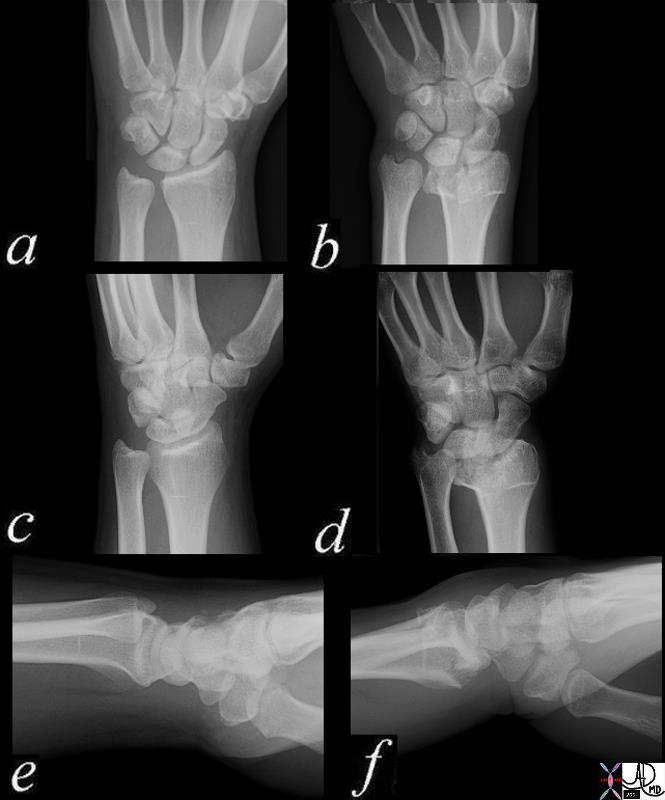
Normal and Colle’s Fracture |
| 70034c03 bone radius ulna wrist dorsally angulated fracture of distal radial metaphysis 2-3 cm proximal to wrist joint,sometimes associated frx of ulnar styloid frx line is almost always on volar side dx Colle’s fracture plain X-ray plain film of wrist normal wrist 70034c03 70034c04 Davidoff MD |
|
Mechanism for Colle’s Fracture |
| 85312p.800 falling outstetched arm Colle’s fracture mechanism of injury bone fracture Davidoff photography Davidoff MD |