
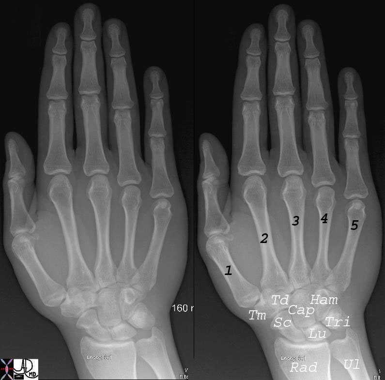
Naming the Digits |
| 45737c02b hand wrist phalanx phalanges radius ulna wrist carpals carpal bones scaphoid lunate triquetrum pisiform hamate hook of hamate capitate tapezoid trapezium metacarpals metacarpal bone sesamoid head body shaft base mip dip pip thumb index middle ring pinky proximal interphalangeal distal interphalangeal middle interphalangeal normal anatomy plain film Courtesy Ashley Davidoff MD 45737 45738 45739 45741 |
The Common Vein
Copyright 2010
Definition
The navicular bone of the hind foot is characterized by being the “keystone of the medial arch”. The navicular links the functions of the hindfoot and forefoot. Its name means little ship.
It is part of the tarsal bones of the foot. The navicular is part of the “acetabulum of the foot”. The “acetabulum of the foot” refers to the structures that support the taluar head including the spring ligament complex and anterior calcaneal facet. It consists of bone and cartilage.
Its unique structural feature is that has 3 sides of cartilage. The shape looks like a boat as it is ovoid and saucer-like. It is located between the talus and cuneiforms. The medial surface has the navicular tuberosity, a site for tendon attachment. The tibialis posterior tendon attaches to the navicular. This medial surface does not contact the ground during weightbearing. The lateral surface comes into contact with the ground during weightbearing.
The blood supply to the navicular is through three zones. Dorsally the dorsalis pedis artery enters the bone. On the plantar surface, the medial plantar artery enters. At the tuberosity, there are branches from the anastomosis between the dorsalis pedis and the medial plantar arteries. The three sites of blood supply do leave a watershed area in the center of the bone that leaves the bone susceptible to avascular necrosis.
The ossification center of the navicular fuses between the 24th to 30th month in boys and between the 18th and 24thmonth in girls. The navicular as well as all other bones, muscles, and ligaments of the body are derived of mesodermal origin in embryo.
The function of the navicular is to link the function of the hindfoot and forefoot through supporting the medial arch. Navicular injuries can have a negative impact on hindfoot motion, gait, and the function of the foot as a unit. The articulation with the talus is very mobile. Articulations with the cuneiforms provide stability and therefore are rigid for medial arch stability.
Common diseases include arthritis, dislocation, fracture, stress fracture, osteomyelitis, coalition, Kohler’s Disease, and accessory navicular.
Arthritis can occur as a result of aging and use (osteoarthritis), rheumatoid arthritis, or as a result of post-traumatic changes.
Subtalar dislocation involves dislocation of the talonavicular and talocalcaneal joints usually as a result of trauma.
Osteomyelitis is an infection of the bone typically due to bacteria.
Calcaneonavicular coalition is an osseous, cartilaginous, or fibrous connection between the two bones. The calcaneonavicular coalition is between the calcaneus and the navicular that usually ossifies between ages 8 to 12 and can cause pain and limited range of motion.
Kohler’s Disease is a self-limiting avascular necrosis of the navicular that presents more commonly in boys between the ages of 5 to 10. Patients have a painful limp and swelling over the navicular bone.
A prominent navicular tuberosity can cause pain when there is compression of the structure by a shoe.
An accessory navicular is an accessory ossicle located on the medial side of the navicular but in continuity with the tibialis posterior tendon. Approximately 2-12% of people have this accessory bone. The accessory navicular fuses at age 9 and may eventually fuse with the navicular.
Commonly used diagnostic procedures include clinical history, physical exam, plain radiographs, CT scan, bone scan, and MRI.
It is usually treated with either non-operative methods or operative methods. Fractures can be treated non-operatively or operatively. Arthritis can be treated with surgery, physical therapy, or NSAIDs. Osteomyelitis is treated with surgery and antibiotics. Coalitions may be treated nonoperatively with cast or surgically by excision of the bony bar or joint fusion. Kohler’s disease is treated with NSAIDs, arch supports, physical therapy, and a short leg walking cast. An accessory navicular or a prominent navicular tuberosity can be treated with cast immobilization or a doughnut shaped padding can be placed over it to relieve pressure. Surgery can be done as well for either an accessory navicular or a prominent navicular tuberosity.
References
Elstrom J, Virkus W, Pankovich (eds), Handbook of Fractures (3rd edition), McGraw Hill, New York, NY, 2006.
Koval K, Zuckerman J (eds), Handbook of Fractures (3rd edition), Lippincott Williams & Wilkins, Philadelphia, PA, 2006.
Lieberman J (ed), AAOS Comprehensive Orthopaedic Review, American Academy of Orthopaedic Surgeons, 2008.
Moore K, Dalley A (eds), Clinically Oriented Anatomy (5th edition), Lippincott Williams & Wilkins, Philadelphia, PA, 2006.
Wheeless’ Textbook of Orthopaedics: Tarsal Navicular (http://www.wheelessonline.com/ortho/tarsal_navicular)