By Gregory R. Waryasz, MD
The Common Vein Copyright 2011
Diagnosis
joint involvement
varus or valgus deformity
stable vs unstable
Plain Film/X-ray Imaging
Plain films are the most basic of all the imaging techniques for musculoskeletal imaging. Bones are well visualized, but there is poor differentiation of soft tissue findings. Fractures are often missed on plain films due to the two-dimensional views. Pattern recognition in the bone can help to generalize bone lesions and give a starting point to determine what the next step should be for the care of the bone lesion. Plain films are often done to monitor bone healing and alignment of fractures to follow the progress.

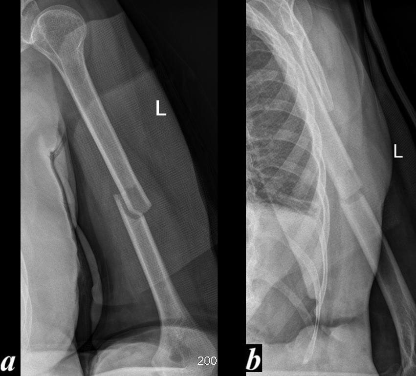
Angulation on the A-P but Aligned on the Lateral The Importance of Two Views |
|
This A-P examination (a) and lateral (b) projection of the left humerus of a 76 year old female following blunt trauma shows an acute almost transverse (with a small oblique component) simple fracture of the middle of the shaft of the left humerus. In the A-P projection the component parts are not touching at all with both components of the fracture being laterally displaced resulting in a varus angulation (red a) . On the lateral examination the fracture is not as well appreciated because the patient could not move her arm and so the chest wall is noted in the background but the component of the fracture are in near anatomic alignment (red b). Courtesy Ashley Davidoff Copyright 2011 103519cL.85 |

Anatomical Alignment Following Closed reduction |
|
This A-P examination of the right humerus of a 76 year old female following blunt trauma shows an acute almost transverse simple fracture of the middle of the shaft of the left humerus. The component parts are not touching at all in this projection with the varus angulation (a). Following closed reduction the component parts are well aligned with significant bone on bone contact and near anatomic alignment. Courtesy Ashley Davidoff 103519cL.8 |
Computed Tomography (CT) Scan
CT scan is a method of imaging that combines two-dimensional x-rays into a larger more detailed picture of the inside of the body. Reformats can be made so that there are images in slices of the axial, coronal, and sagittal planes of the body. CT scan is important for following subtle fractures and further classifying injuries. CT scans are better than x-ray at visualizing soft tissue structures. CT scans are mainstay in trauma to visualize the brain, chest, abdomen, and pelvis. Current technology allows the CT scans to be made into three dimensional reconstructions that can help with surgical planning. The downside to CT scan is the large amount of radiation exposure.
MRI is a method of imaging that uses magnets to help make images utilizing the hydrogen atoms in water in the body rather than ionizing radiation. MRI is the best modality for soft tissue injuries including ligaments, tendons, and muscles. Bone edema can be an indication of a fracture that could not be visualized on CT or plain film. MRI is very important in sports medicine. MRI uses different sequences to help the radiologist make a diagnosis. MRI is a good modality for evaluating bone for osteomyelitis.

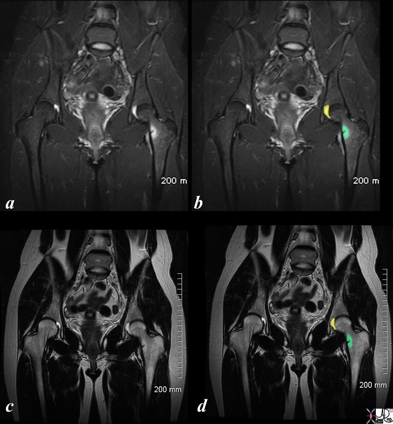
82987cL01s.8 |
|
These MRI images demonstrate a stress fracture in the neck of the left femur. Images a and b are STIR images in the coronal plane while images c and d show a T2 sequence in the same plane. The fracture is characterized by a small medial positioned fracture (black) surrounded by a rim of edema (green in b and d). A small effusion is noted in the left hip (yellow) Courtesy Ashley Davidoff Copyright 2011 82987cL01s.8 |
Ultrasound
Ultrasound uses sound waves to generate an image. Although primarily used for vascular, gynecologic, and internal organ examination, ultrasound is being used to evaluate soft tissue structures of the appendicular skeleton and the joint spaces. Many pediatric rheumatologists use ultrasound to look for effusions as this modality is safer for children that other modes of imaging. Ultrasound can also be used for interventional procedures such as biopsies and aspirations.
Bone Scan
Bone scan is a nuclear medicine test that helps to look for activity in bone consistent with increased metabolism. Bone scan requires an injection of radioactive material such as technetium-99m-MDP to help isolate areas of interest. Conditions that can use bone scan for evaluation include looking for metastatic lesions, evaluating bone lesions, and finding subtle fractures. Bone scan usually requires correlation with other imaging modalities.

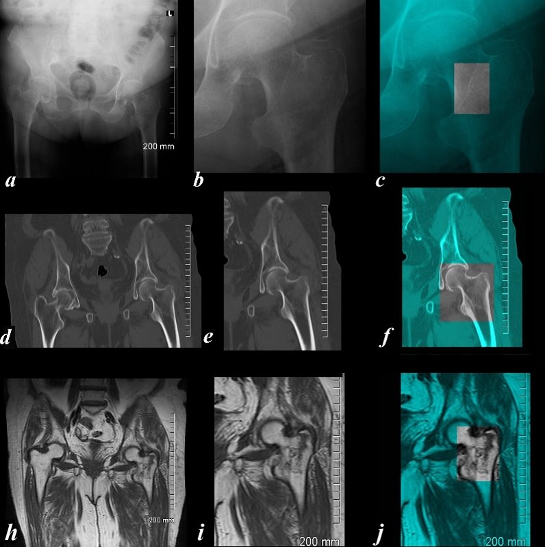
Plain Film, CTscan and MRI of an Non Displaced Intertrochanteric Fracture |
|
The A-P examination of the proximal left femur of an 86 year old female shows the three major technologies in a difficult to diagnose fracture. Images a,b and c show a subtle change(?break) in the intertrochanteric line best seen in image c which is overlaid in teal green, and the area of interest shown in black and white. Image d,e,and f show a sagittally reformatted CT scan where there is a failure to visualize any hint of a fracture and repeated review of the axial images were never convincing for a fracture. Because the pain was severe and incapacitating an MRI was performed and the T1 component of that examination is shown in images (g,h,i) revealing an obvious non displaced intertrochanteric fracture of the left femur best visualized in i by a ragged intertrochanteric black line. CT is usually a quick and highly sensitive study for fractures but in rare instances such as this case, when the patient’s history creates ongoing concern, MRI proved to be highly sensitive to the injury. Courtesy Ashley Davidoff Copyright 2011 103778c03L.8 |
References
Elstrom J, Virkus W, Pankovich (eds), Handbook of Fractures (3rd edition), McGraw Hill, New York, NY, 2006.
Koval K, Zuckerman J (eds), Handbook of Fractures (3rd edition), Lippincott Williams & Wilkins, Philadelphia, PA, 2006.
Lieberman J (ed), AAOS Comprehensive Orthopaedic Review, American Academy of Orthopaedic Surgeons, 2008.
